Knee Muscle Anatomy Mri - Medical imaging and radiological anatomy : X-ray, CT, MRI ... / Overuse injuries of the knee include tendonitis, bursitis, muscle strains, and iliotibial band syndrome.
Knee Muscle Anatomy Mri - Medical imaging and radiological anatomy : X-ray, CT, MRI ... / Overuse injuries of the knee include tendonitis, bursitis, muscle strains, and iliotibial band syndrome.. Radiology imaging medical anatomy human anatomy and physiology anatomy study. 4, infrapatellar fat pad of hoffa. Find out how the different structures fit together in our knee diagram the knee joint is the largest and one of the most complex joints in the human body. Each anatomical structure was labeled interactively. The muscles of the knee include the quadriceps, hamstrings, and the muscles of the calf.
This mri knee cross sectional anatomy tool is absolutely free to use. Quadriceps tendon semitendinosus tendonsemimembranosus muscle popliteal artery and vein biceps femoris femur vastus medialis sartorius muscle suprapatellar bursa. An understanding of normal anatomy and biomechanics of the knee extensor mechanism is necessary to comprehend the imaging of extensor mechanism injuries. Injuries of the patellofemoral joint. Magnetic resonance imaging (mri scan):
Anterior Muscles of Knee Stock Photo: 7710697 - Alamy from c8.alamy.com Functional anatomy of the shoulder complex malcolm peat the shoulder complex, together with other joint and muscle mechanisms of the upper limb. Song, uc san francisco msiv gillian lieberman md. General anatomy and musculoskeletal system. Use the checklist to quiz yourself. Find out how the different structures fit together in our knee diagram the knee joint is the largest and one of the most complex joints in the human body. Want to learn more about it? Involved early gray = muscle: Please email baodo at stanford.edu.
Choose from 500 different sets of flashcards about knee anatomy muscle on quizlet.
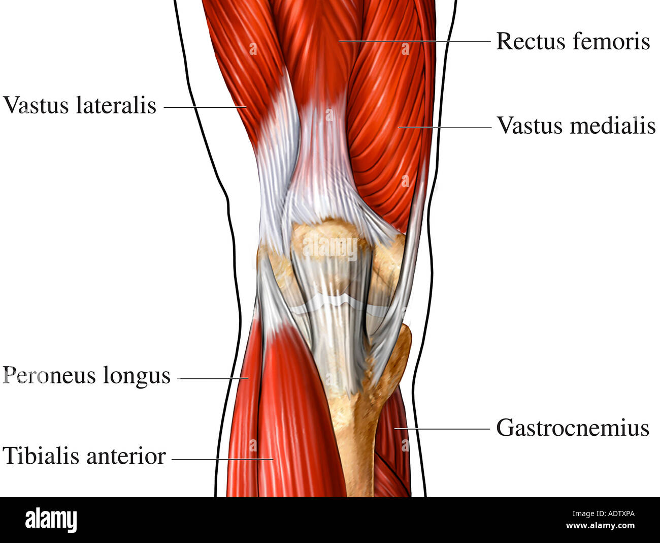
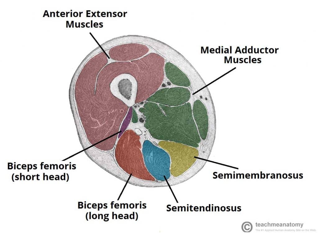
This section of the website will explain large and minute details of sagittal knee cross sectional anatomy. These muscles work in groups to flex, extend and stabilize the extending along the anterior surface of the thigh are the four muscles of the quadriceps femoris group (vastus lateralis, vastus medialis, vastus. Knee anatomy francesc malagelada jordi vega pau golanó the knee is the largest joint in the human body and one of the most complex from a functional point of view. 4, infrapatellar fat pad of hoffa. Involved early gray = muscle: Find out how the different structures fit together in our knee diagram the knee joint is the largest and one of the most complex joints in the human body. 12 photos of the knee muscle anatomy mri. Mr imaging of knees having isolated and combined ligament injuries. This webpage presents the anatomical structures found on knee mri. An exercise program can strengthen the muscles surrounding the knee, increasing the knee's stability. Musculoskeletal radiology south texas radiology group. Click now to learn more about the bones, muscles, and soft tissues of these regions at leg and knee anatomy: It is also one of the most often injured joints because of its anatomic characteristics, the interrelation of its structural components.
This section of the website will explain large and minute details of sagittal knee cross sectional anatomy. Click on the links to show each structure. An understanding of normal anatomy and biomechanics of the knee extensor mechanism is necessary to comprehend the imaging of extensor mechanism injuries. Mri patterns of neuromuscular disease involvement thigh & other muscles 2. It is constructed by 4 bones and an extensive network of ligaments and muscles.1.
Muscles of the Posterior Thigh - Hamstrings - Damage ... from teachmeanatomy.info Radiology imaging medical anatomy human anatomy and physiology anatomy study. 12 photos of the knee muscle anatomy mri. Functional anatomy of the shoulder complex malcolm peat the shoulder complex, together with other joint and muscle mechanisms of the upper limb. Learn anatomy using a full pacs! Magnetic resonance imaging (mri scan): Each anatomical structure was labeled interactively. Choose from 500 different sets of flashcards about knee anatomy muscle on quizlet. Mri for evaluating knee pain in older patients:
The muscles that affect the knee's movement run along the thigh and calf.
It is constructed by 4 bones and an extensive network of ligaments and muscles.1. Mr imaging of knees having isolated and combined ligament injuries. Song, uc san francisco msiv gillian lieberman md. Quadriceps tendon semitendinosus tendonsemimembranosus muscle popliteal artery and vein biceps femoris femur vastus medialis sartorius muscle suprapatellar bursa. Involved early gray = muscle: Use the mouse to scroll or the arrows. This mri knee cross sectional anatomy tool is absolutely free to use. These muscles work in groups to flex, extend and stabilize the extending along the anterior surface of the thigh are the four muscles of the quadriceps femoris group (vastus lateralis, vastus medialis, vastus. This webpage presents the anatomical structures found on knee mri. Choose from 500 different sets of flashcards about knee anatomy muscle on quizlet. Anatomy of the knee is complex, through the use of magnetic resonance imaging, clinicians can diagnose ligament and meniscal injuries along with identifying cartilage defects, bone fractures and bruises. 12 photos of the knee muscle anatomy mri. Mr arthrogram knee loose osteochondral lesion.
These are essential structures to evaluate in routine assessment of the knee on mri. Please email baodo at stanford.edu. There are various muscles that control movement, ligaments that. Learn anatomy using a full pacs! Song, uc san francisco msiv gillian lieberman md.
Knee Mri Anatomy Related Keywords & Suggestions - Knee Mri ... from i.pinimg.com The muscles that affect the knee's movement run along the thigh and calf. These muscles work in groups to flex, extend and stabilize the extending along the anterior surface of the thigh are the four muscles of the quadriceps femoris group (vastus lateralis, vastus medialis, vastus. Injuries of the patellofemoral joint. Involved early gray = muscle: Knee anatomy francesc malagelada jordi vega pau golanó the knee is the largest joint in the human body and one of the most complex from a functional point of view. Learn anatomy using a full pacs! Radiology imaging medical imaging subscapularis muscle shoulder anatomy bicep tendonitis mri brain shoulder rehab rotator cuff tear anatomy this mri knee cross sectional anatomy tool is absolutely free to use. Please email baodo at stanford.edu.
Learn anatomy using a full pacs!
Song, uc san francisco msiv gillian lieberman md. 12 photos of the knee muscle anatomy mri. This webpage presents the anatomical structures found on knee mri. It is constructed by 4 bones and an extensive network of ligaments and muscles.1. Free access interactive and dynamic anatomical atlas. Mr arthrogram knee loose osteochondral lesion. Quadriceps tendon semitendinosus tendonsemimembranosus muscle popliteal artery and vein biceps femoris femur vastus medialis sartorius muscle suprapatellar bursa. Want to learn more about it? They are attached to the femur (thighbone), tibia (shinbone), and fibula (calf bone) by fibrous tissues called ligaments. Anatomy of peritoneum and mesentery. Magnetic resonance imaging (mri scan): Knee joint anatomy is complex with muscles, ligaments, cartilage and tendons. There are various muscles that control movement, ligaments that.